Skin Whitening Mechanism Diagram
Create a paper-ready skin whitening mechanism diagram showing UV signaling, α-MSH, MC1R, MITF, tyrosinase, and melanin synthesis inhibition. Free prompt template for papers and PPT.
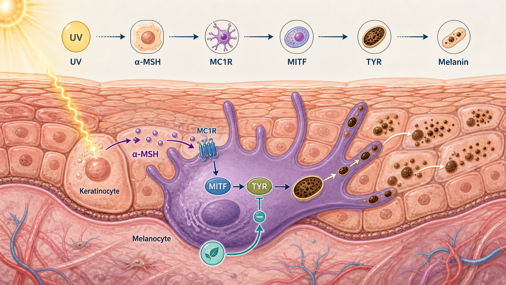
这张机制图展示了什么
A useful skin whitening mechanism diagram does more than list ingredients. It shows how UV and inflammatory cues drive melanogenesis, which nodes (MC1R → MITF → tyrosinase) control melanin production, and where common brightening actives intervene—so a reviewer or client can follow the story in one figure.
- 1UV / ROS or α-MSH acts as the upstream trigger on melanocytes (and optionally keratinocyte signals).
- 2MC1R signaling raises MITF activity, which turns on tyrosinase and melanogenic enzymes.
- 3Melanin is produced and packaged into melanosomes.
- 4Melanosomes transfer toward keratinocytes, changing visible pigmentation.
- 5Brightening actives are annotated at enzyme inhibition, signaling dampening, or transfer reduction points.
What searchers and reviewers expect in this figure
People searching for a skin whitening mechanism diagram usually need a slide- or paper-ready schematic: clear left-to-right or top-to-bottom flow, short node labels, and an endpoint such as reduced melanin synthesis or reduced melanosome transfer—not a decorative illustration with no pathway logic.
Strong figures separate (1) the physiological cascade and (2) intervention points. That makes the same base diagram reusable for niacinamide, tranexamic acid, arbutin, vitamin C derivatives, or a multi-active claim without redrawing the whole cascade.
Core pathway nodes to include
Start from UV / ROS or α-MSH as the input. Connect MC1R on melanocytes to MITF, then to tyrosinase and related enzymes (TYRP1/TYRP2 when space allows). End with melanin packaging and transfer toward keratinocytes if your claim is “brighter appearance,” not only enzyme inhibition.
Optional but high-value callouts: keratinocyte-derived SCF/ET-1, inflammation crosstalk, and a small “intervention” row under the cascade. Keep labels short enough to stay legible at 16:9 PPT size.
How to generate your version in MechFig
Open the reusable prompt below, replace the active name and intervention point, then generate. Prefer a horizontal pathway or graphical-abstract layout for manuscripts; use a simpler slide style for brand decks.
If you need a related cosmetic science figure next, continue to barrier repair or retinol mechanism pages and keep visual style consistent across the deck.
可复用提示词
将提示词放入 MechFig,再把成分、通路或目标组织替换为你的研究内容。
Scientific mechanism infographic, clean flat vector style, labeled arrows between steps, soft blue-purple academic palette, white background, suitable for research paper or PPT, Chinese and English labels ok, high clarity, no photorealistic skin texture. Scientific content: UV triggers keratinocytes to release α-MSH → binds MC1R on melanocytes → cAMP/PKA → MITF upregulation → tyrosinase (TYR) expression → tyrosine to dopaquinone → melanin. Whitening actives inhibit TYR or block α-MSH signaling.
常见问题
What should a skin whitening mechanism diagram include?
Include the trigger (UV/α-MSH), key nodes (MC1R, MITF, tyrosinase), the melanin endpoint, and clearly marked intervention points for your active. Use directional arrows and short labels so the figure works in both papers and PPT.
Is this only for cosmetics marketing?
No. The same melanogenesis cascade is used in cosmetic science, dermatology education, and research graphical abstracts. Change the intervention layer to match your claim strength and evidence level.
Can I generate this figure with AI in MechFig?
Yes. Use the prompt on this page, swap in your ingredient and target node, and generate a pathway-style figure. Iterate labels until the cascade matches your manuscript or slide narrative.