Blood-Brain Barrier Diagram
Explore a blood-brain barrier diagram with brain endothelial tight junctions, pericytes, astrocyte endfeet, transporters, and selective permeability.
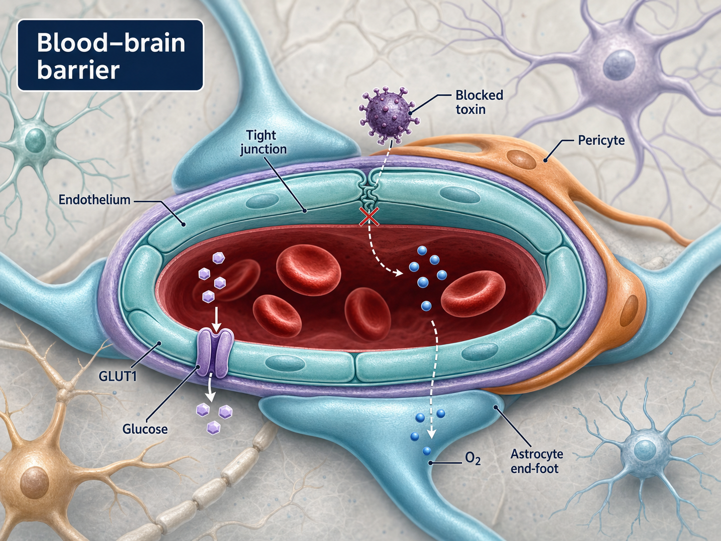
What this mechanism diagram shows
This blood-brain barrier diagram shows the neurovascular unit as a coordinated interface rather than a single vessel wall. Brain microvascular endothelial cells and tight junctions form the primary barrier, while pericytes, astrocyte endfeet, basement membrane, transporters, and efflux pumps help regulate selective movement between blood and neural tissue.
- 1Brain microvascular endothelial cells form the vessel wall and minimize non-selective passage.
- 2Tight junction complexes restrict paracellular diffusion between endothelial cells.
- 3Pericytes, basement membrane, and astrocyte endfeet support barrier phenotype and signaling.
- 4Nutrient transporters and selected transcytosis routes regulate controlled entry into brain tissue.
- 5Efflux pumps return many xenobiotics toward the blood and limit brain exposure.
Anatomy to include in a blood-brain barrier diagram
A useful cross-section should identify the vessel lumen, endothelial cells, tight junctions, basement membrane, pericytes, astrocyte endfeet, and the brain parenchyma. These elements explain why the blood-brain barrier is a multicellular neurovascular unit rather than a generic capillary wall.
When space permits, label representative tight-junction proteins such as claudin-5, occludin, and ZO-1. Keep cellular geometry simple enough that transport routes and permeability changes remain legible.
Transport routes and barrier mechanisms
Show passive diffusion for selected small lipophilic molecules, carrier-mediated transport for nutrients, receptor-mediated transcytosis where relevant, and efflux pumps such as P-glycoprotein. Paracellular movement should be visibly restricted by tight junctions in the intact-barrier state.
For disease or drug-delivery figures, use a second panel to show barrier disruption, inflammatory signaling, altered tight junctions, or a delivery strategy. Separating normal and altered states prevents contradictory arrows from crowding one vessel cross-section.
Generate a custom BBB research figure
Use the prompt below and specify whether the figure is anatomical, mechanistic, or focused on drug delivery. Name the transport route, cell type, target molecule, and desired endpoint so the generated image does not substitute generic brain imagery for a real barrier schematic.
Check anatomical orientation, transporter direction, cell labels, and the evidence for any claimed permeability change before publication. For quantitative studies, keep measurements and statistical results outside the illustrative mechanism panel.
Scientific sources and content method
Sources checkedThis mechanism text was prepared with an AI-assisted editorial workflow and checked against the peer-reviewed sources below. It is for research communication and education, not medical advice; verify it against your study context before publication.
Last source check:
- The Blood–Brain Barrier
Cold Spring Harbor Perspectives in Biology (2015) · DOI: 10.1101/cshperspect.a020412
Reusable prompt
Paste this into MechFig, then replace the ingredient, pathway, or target tissue with your own research context.
Blood-brain barrier cross-section scientific diagram, labeled cells, academic palette. Scientific content: Brain endothelium tight junctions + pericytes/astrocyte endfeet → selective permeability; transporters and efflux pumps control entry.
Embed this mechanism diagram
Copy the HTML below to include descriptive image text and attribution back to this example page. Confirm that your use complies with applicable rights and site terms before publication or commercial use.
FAQ
What should a blood-brain barrier diagram include?
Include endothelial cells, tight junctions, basement membrane, pericytes, astrocyte endfeet, blood and brain compartments, plus the transport routes relevant to your topic.
Is the blood-brain barrier just tight junctions?
No. Tight junctions are central, but transporters, efflux pumps, endothelial properties, pericytes, astrocytes, and basement membrane all contribute to barrier function.
Can this template illustrate drug delivery across the BBB?
Yes. Add the therapeutic cargo, its transport mechanism, the endothelial route, and the brain-side destination. Distinguish validated transport from a proposed mechanism.