MAPK/ERK Pathway Diagram
Explore a MAPK/ERK pathway diagram from growth-factor receptor activation through Ras, Raf, MEK, ERK, nuclear signaling, and cellular outcomes.
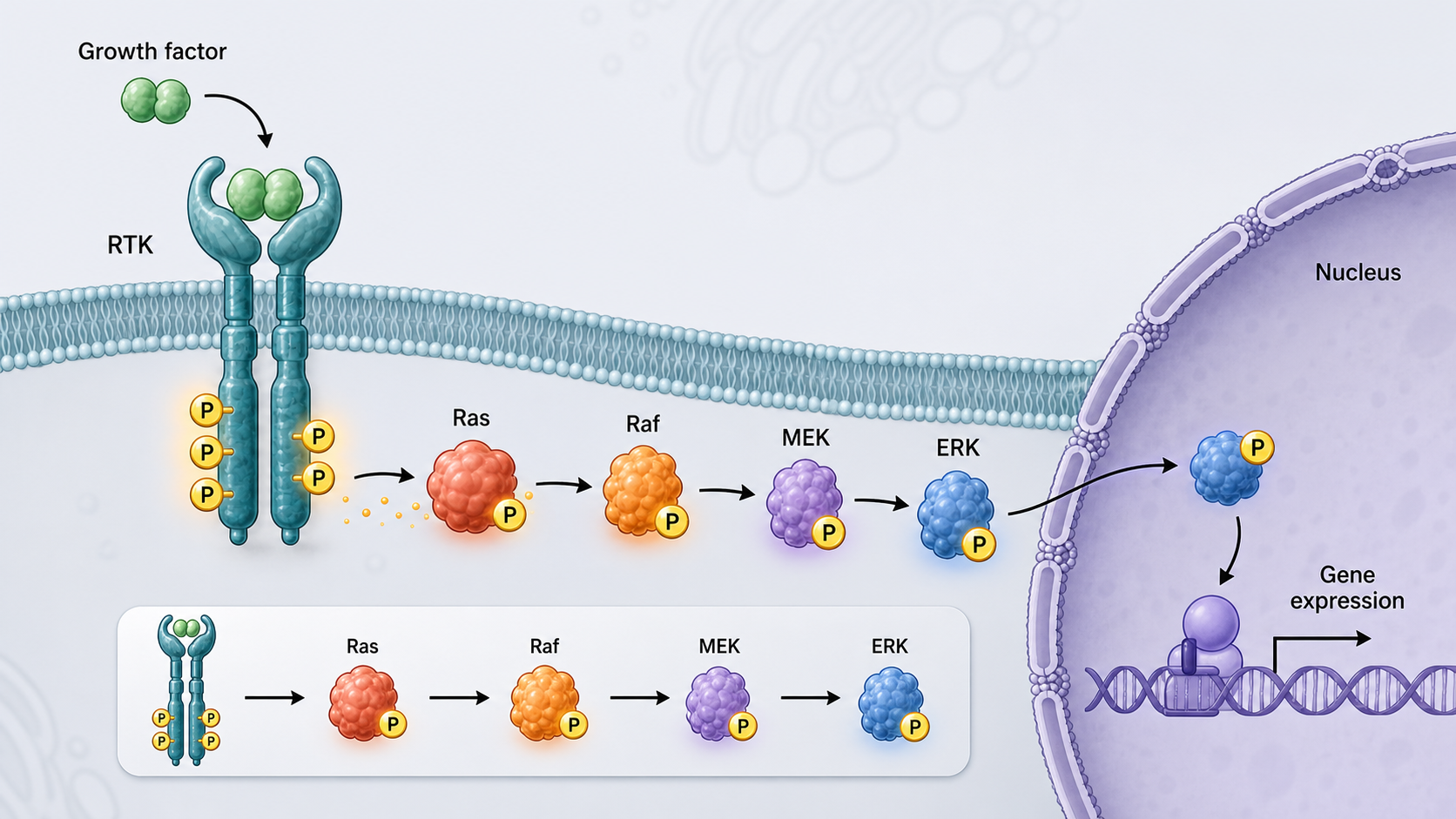
What this mechanism diagram shows
The canonical MAPK/ERK cascade carries signals from activated receptor tyrosine kinases through Grb2/SOS, Ras, Raf, MEK, and ERK. Activated ERK regulates cytoplasmic substrates and enters the nucleus to influence transcription, proliferation, differentiation, and survival in a context-dependent manner.
- 1A growth factor activates a receptor tyrosine kinase at the plasma membrane.
- 2Grb2 and SOS promote conversion of Ras-GDP to active Ras-GTP.
- 3Ras recruits and activates Raf, which phosphorylates MEK.
- 4MEK phosphorylates ERK, enabling cytoplasmic and nuclear signaling.
- 5ERK regulates context-dependent transcriptional and cellular responses.
Core MAPK/ERK pathway nodes
Start at a growth factor and receptor tyrosine kinase, then show Grb2/SOS-mediated Ras activation followed by the Raf–MEK–ERK kinase cascade. Separate membrane, cytoplasm, and nucleus so location changes remain readable.
Use phosphorylation labels selectively and place inhibitors beside their actual targets. Feedback regulators such as DUSPs or Sprouty can be added when the research question concerns signal duration or resistance.
How to adapt the diagram for research
Cancer figures may add activating RAS or BRAF variants and targeted inhibitors; developmental figures may emphasize signal strength and duration. Keep the central cascade visually dominant and add only evidence-supported branches.
Distinguish pathway activation from a guaranteed phenotype. ERK output depends on cell type, timing, intensity, and crosstalk with other pathways.
Generate a custom MAPK pathway figure
Use the prompt below, specify the receptor, mutation, inhibitor, and endpoint, then verify protein names, phosphorylation direction, and cellular compartment before publication.
Scientific sources and content method
Sources checkedThis mechanism text was prepared with an AI-assisted editorial workflow and checked against the peer-reviewed sources below. It is for research communication and education, not medical advice; verify it against your study context before publication.
Last source check:
- ERK signalling: a master regulator of cell behaviour, life and fate
Nature Reviews Molecular Cell Biology (2020) · DOI: 10.1038/s41580-020-0255-7
Reusable prompt
Paste this into MechFig, then replace the ingredient, pathway, or target tissue with your own research context.
Classic cell signaling cascade infographic, vertical or horizontal kinase cascade, protein nodes with short labels, blue-gray scientific palette, white background, paper-ready. Scientific content: GF binds RTK → dimerization & autophosphorylation → Grb2-Sos → Ras-GTP → Raf → MEK → ERK → nuclear TFs (c-Fos, c-Myc) → proliferation genes.
Embed this mechanism diagram
Copy the HTML below to include descriptive image text and attribution back to this example page. Confirm that your use complies with applicable rights and site terms before publication or commercial use.
FAQ
What should a MAPK/ERK pathway diagram include?
Include the receptor, Grb2/SOS, Ras, Raf, MEK, ERK, cellular compartments, and a qualified downstream output. Add mutations or inhibitors only when relevant.
Are MAPK and ERK the same thing?
ERK1/2 are MAP kinases within the Ras–Raf–MEK–ERK pathway. MAPK is a broader family that also includes pathways such as JNK and p38.
How should MAPK inhibitors be shown?
Place each inhibitor at its direct target, such as BRAF or MEK, and use an inhibitory connector rather than a detached compound list.