NF-κB Pathway Diagram
Explore a canonical NF-κB pathway diagram showing receptor signaling, IKK activation, IκB degradation, p65/p50 nuclear entry, and inflammatory gene expression.
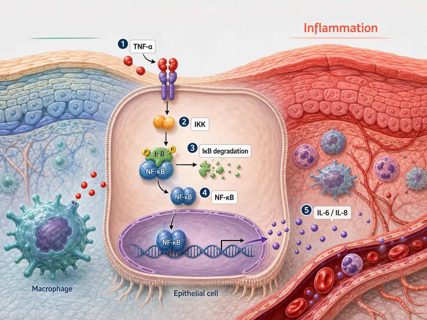
What this mechanism diagram shows
In canonical NF-κB signaling, stimuli such as TNF or microbial products activate receptor-proximal complexes and the IKK complex. IKK phosphorylates IκB, leading to its ubiquitination and degradation. Released p65/p50 dimers enter the nucleus and regulate context-dependent inflammatory, immune, survival, and feedback genes.
- 1TNF, a microbial cue, or another stimulus activates its cell-surface receptor.
- 2Receptor-proximal signaling activates the IKK complex.
- 3IKK phosphorylates IκB, marking it for ubiquitination and proteasomal degradation.
- 4Released p65/p50 NF-κB translocates into the nucleus.
- 5NF-κB regulates context-dependent inflammatory and feedback genes.
Structure of a canonical NF-κB figure
Show an upstream stimulus and receptor, the receptor-proximal signaling complex, IKK, IκB-bound NF-κB, proteasomal IκB degradation, and nuclear p65/p50. Separating resting and activated states makes the release step easy to understand.
If the topic is TNF signaling, label TNFR-associated components; for TLR signaling, use the appropriate adaptor path. Do not merge distinct receptor branches into one unlabeled arrow.
Inflammatory outputs and negative feedback
Representative outputs may include cytokines, chemokines, adhesion molecules, and IκBα resynthesis. Select a small gene set that matches the tissue and experiment rather than implying that every NF-κB target changes in every cell.
The non-canonical pathway uses different upstream machinery and RelB/p52 output. Label the page as canonical when that is the scope.
Generate an NF-κB mechanism diagram
Specify the stimulus, receptor branch, cell type, intervention, and measured endpoint in the prompt. Verify adaptor proteins and inhibitor targets before using the generated figure in a manuscript.
Scientific sources and content method
Sources checkedThis mechanism text was prepared with an AI-assisted editorial workflow and checked against the peer-reviewed sources below. It is for research communication and education, not medical advice; verify it against your study context before publication.
Last source check:
- Shared Principles in NF-κB Signaling
Cell (2008) · DOI: 10.1016/j.cell.2008.01.020
Reusable prompt
Paste this into MechFig, then replace the ingredient, pathway, or target tissue with your own research context.
Intracellular signaling pathway diagram, cytoplasm to nucleus, clear inhibition and activation arrows, academic textbook style, high contrast labels. Scientific content: TNF-α/IL-1 → receptor → IKK activation → IκB phosphorylation & proteasomal degradation → NF-κB (p65/p50) nuclear translocation → inflammatory genes (COX-2, IL-6, TNF).
Embed this mechanism diagram
Copy the HTML below to include descriptive image text and attribution back to this example page. Confirm that your use complies with applicable rights and site terms before publication or commercial use.
FAQ
What should an NF-κB pathway diagram include?
For the canonical pathway include the stimulus, receptor, IKK complex, IκB, p65/p50, proteasome, nucleus, and selected target genes.
What is the difference between canonical and non-canonical NF-κB signaling?
Canonical signaling commonly uses IKKβ-dependent IκB degradation and p65/p50. Non-canonical signaling centers on NIK, IKKα, p100 processing, and RelB/p52.
Can I add an anti-inflammatory compound?
Yes, but position it at the experimentally supported target and distinguish direct inhibition from a downstream reduction in inflammatory markers.