Wnt/β-Catenin Pathway Diagram
Explore a Wnt/β-catenin pathway diagram showing Frizzled/LRP, the destruction complex, β-catenin stabilization, nuclear entry, and TCF/LEF transcription.
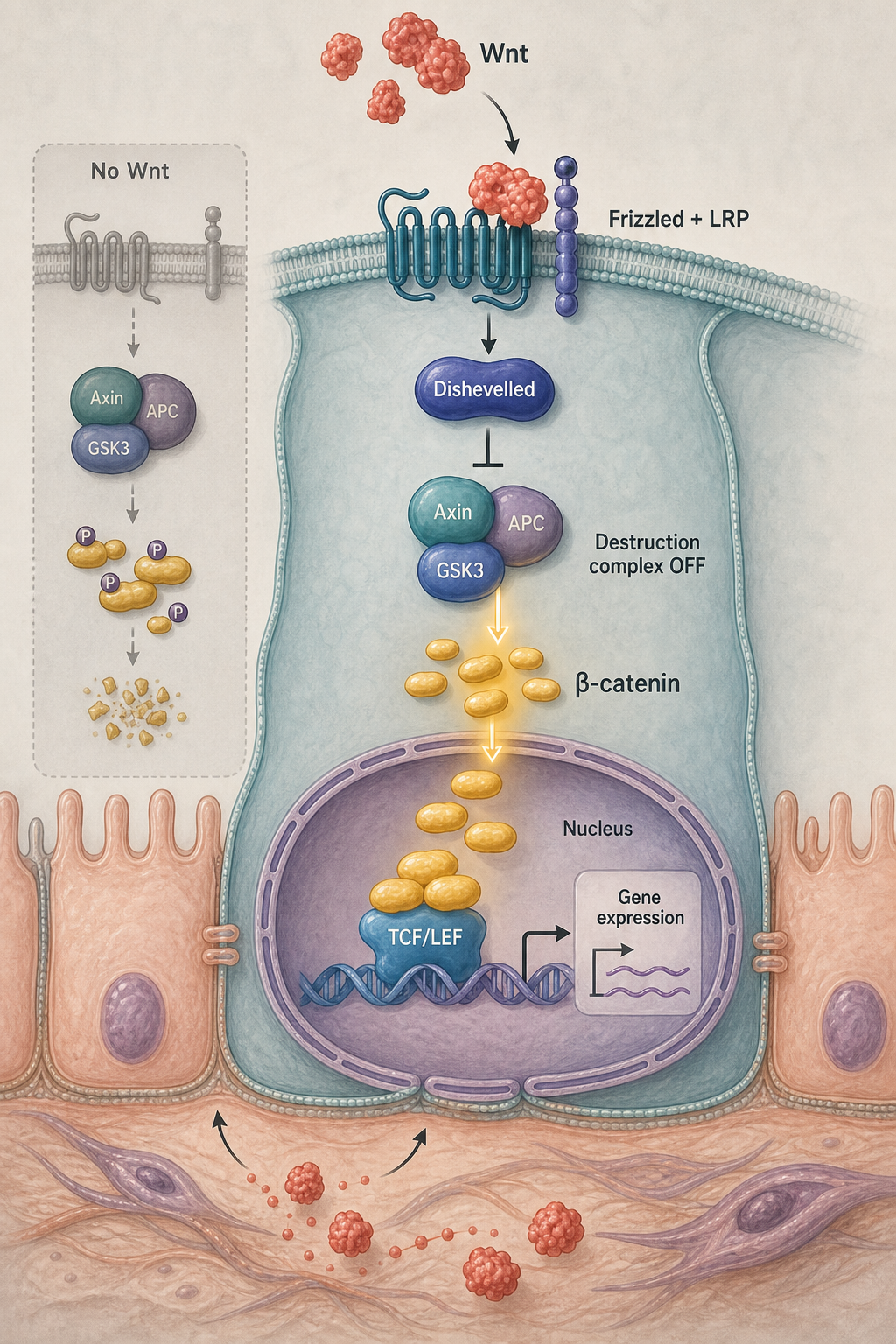
What this mechanism diagram shows
This Wnt/β-catenin pathway diagram compares the pathway with and without Wnt ligand. In the OFF state, the Axin–APC–GSK3β–CK1 destruction complex phosphorylates β-catenin for degradation. In the ON state, Wnt binds Frizzled and LRP5/6, Dishevelled suppresses destruction-complex activity, and stabilized β-catenin enters the nucleus to regulate TCF/LEF target genes.
- 1Without Wnt, the Axin–APC–GSK3β–CK1 destruction complex phosphorylates β-catenin.
- 2Phosphorylated β-catenin is ubiquitinated and degraded, keeping TCF/LEF target genes repressed.
- 3Wnt binds Frizzled and LRP5/6 and recruits Dishevelled at the membrane.
- 4Destruction-complex activity falls, allowing β-catenin to accumulate and enter the nucleus.
- 5β-catenin partners with TCF/LEF to regulate context-dependent target genes.
How to structure a canonical Wnt pathway figure
A useful Wnt pathway figure should make the OFF and ON states visually distinct. The OFF panel explains why cytosolic β-catenin remains low; the ON panel shows ligand–receptor binding, β-catenin stabilization, nuclear translocation, and transcriptional output. This side-by-side structure is easier to read than one crowded cascade.
Keep the membrane, cytoplasm, and nucleus visually separated. Use activation arrows for Wnt–Frizzled/LRP signaling and an inhibitory mark where Dishevelled disrupts the destruction complex. Label ubiquitination or proteasomal degradation only when it supports the teaching or research question.
Core Wnt/β-catenin nodes to include
The minimum canonical pathway includes Wnt ligand, Frizzled, LRP5/6, Dishevelled, the Axin/APC/GSK3β/CK1 destruction complex, β-catenin, and TCF/LEF. Optional outputs include proliferation, differentiation, stem-cell maintenance, or a disease-specific target gene set.
If the figure discusses inhibitors or therapeutics, place each intervention beside its actual target rather than collecting compounds in a detached legend. That makes the mechanism of action readable at manuscript and presentation size.
Generate a custom Wnt pathway diagram
Use the reusable prompt below as a starting structure, then specify whether you need the canonical pathway, a disease context, or crosstalk with PI3K/AKT, Hippo/YAP, or TGF-β signaling. Ask for a horizontal two-state layout when comparison is the priority.
Treat the generated image as a scientific draft. Verify protein names, arrow direction, compartment placement, and the evidence behind any claimed downstream phenotype before using the figure in a paper or teaching material.
Scientific sources and content method
Sources checkedThis mechanism text was prepared with an AI-assisted editorial workflow and checked against the peer-reviewed sources below. It is for research communication and education, not medical advice; verify it against your study context before publication.
Last source check:
- Wnt/β-Catenin Signaling and Disease
Cell (2012) · DOI: 10.1016/j.cell.2012.05.012
Reusable prompt
Paste this into MechFig, then replace the ingredient, pathway, or target tissue with your own research context.
Wnt beta-catenin pathway with and without ligand, clean cell signaling figure. Scientific content: Wnt → Frizzled/LRP → destruction complex off → β-catenin nuclear → TCF/LEF transcription.
Embed this mechanism diagram
Copy the HTML below to include descriptive image text and attribution back to this example page. Confirm that your use complies with applicable rights and site terms before publication or commercial use.
FAQ
What should a Wnt/β-catenin pathway diagram include?
Include Wnt, Frizzled/LRP5/6, Dishevelled, the Axin/APC/GSK3β/CK1 destruction complex, β-catenin, the nucleus, and TCF/LEF-mediated transcription. A two-panel OFF-versus-ON layout is usually clearest.
Is Wnt signaling the same as the β-catenin pathway?
Canonical Wnt signaling is β-catenin dependent. Wnt ligands can also activate non-canonical pathways, so label the figure “canonical Wnt/β-catenin” when that is the intended scope.
Can I adapt this diagram for cancer or stem-cell research?
Yes. Replace the generic endpoint with evidence-supported genes or phenotypes and add disease-specific mutations or inhibitors at the correct pathway node.